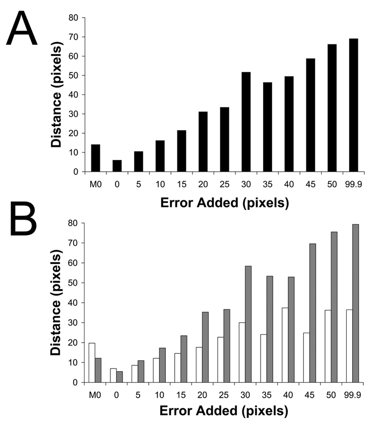
Figure 4.
Error in 2D spot placement affects the accuracy of orientation determination. A 5fold-symmetric reference consisting of native virus and a single 3D dot was used to orient 2D images in which various levels of error were applied to the spot coordinates. The reference dot was rotated according to the resulting Eulerian angles, projected in 2D, and its Cartesian coordinates were compared with the true dot position for each image. The RMS coordinate difference is reported in pixels for error levels of 0, 5, 10, 15, 20, 25, 30, 35, 40, 45, 50, and 99.9 pixels. The leftmost column (M0) refers to dots placed manually in Bubeck et al (2005b). Orientation accuracy for all dotted images is reported in panel A. Images were classified according to the distance from the center of the virus to the spot position (B). Grey bars represent distances more than 76.9 pixels radius, while white bars illustrate those less than 76.9 pixels. Manual spot placement was more accurate for high-radius particles where density for receptor molecules was more often visible.

