
Figure 7.
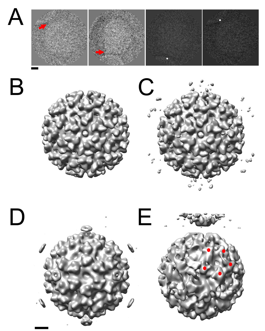
The post-imaging fiducial marker method was used to generate 3D reconstructions of SFV that provide the first experimental evidence that the virus initiates fusion along its 5fold axis. (A) The membrane contact point in the 2D images was identified (red arrows, left two panels) and artificially dotted (right two panels). (B) An icosahedral reconstruction calculated from unmodified images demonstrates that the viral component of the complex remains similar overall to the previously published structure that lacks membrane. (C) The corresponding reconstruction calculated from dotted images shows weak density features (black arrow) encircling the viral 5fold axes 373 Å from the center of the virus. (D). This density was enhanced (black arrow) when only those particles that were visually confirmed to be interacting with the membrane (A, right two panels) were included. (E) The optimized method successfully generated a preliminary 5fold-symmetric model of the SFV-liposome complex to be used in further bootstrap orientation refinement. Red spheres indicate the positions of 5 spikes on the viral surface that are related by an icosahedral symmetry operator that was not enforced in the reconstruction. Scale bars: 100 Å