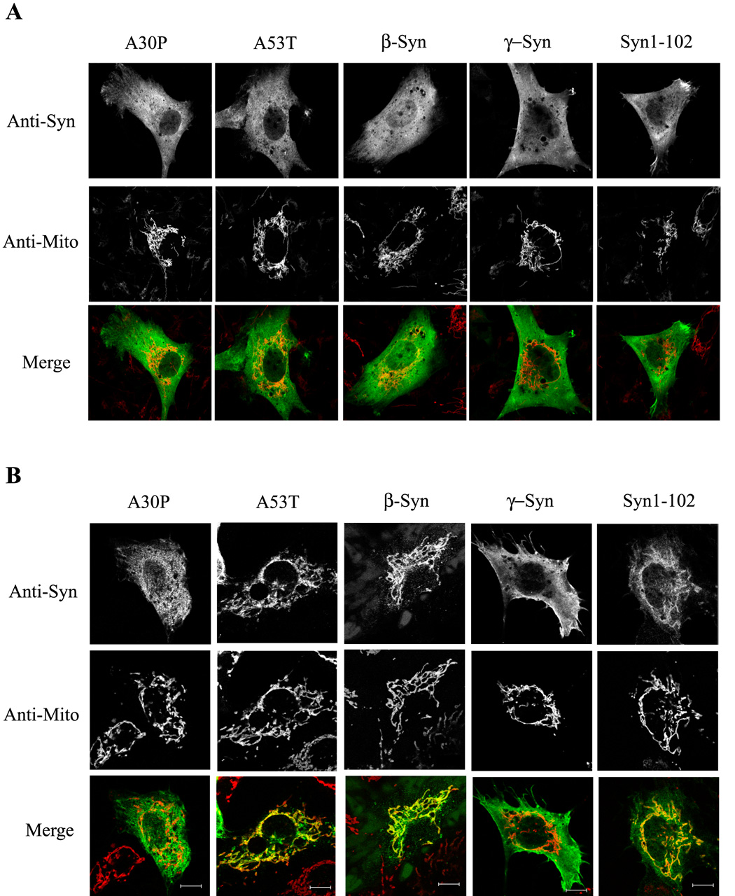
Figure 2. Effects of oxidative stress on translocation of different synucleins to mitochondria.

(A) Representative immunofluorescence images of SK-N-SH cells transiently expressing various synucleins. Cells were untreated (A) or treated with 250 µM H2O2 for 2 h (B), before being fixed as in Fig. 1. Note that A53T, β-synuclein, and Syn1-102 translocate to mitochondria with H2O2, whereas translocation was minimal with A30P and γ-synuclein. Mitochondria were labeled either with antibodies to the outer mitochondrial membrane protein Tom20 or to the intermembrane space protein cytochrome c, depending on the species of primary synuclein antibody used. Cells expressing low levels of various synucleins are depicted. Bar: 10 µm.
