
Figure 4. Synuclein translocates to mitochondria during acidic fixation.
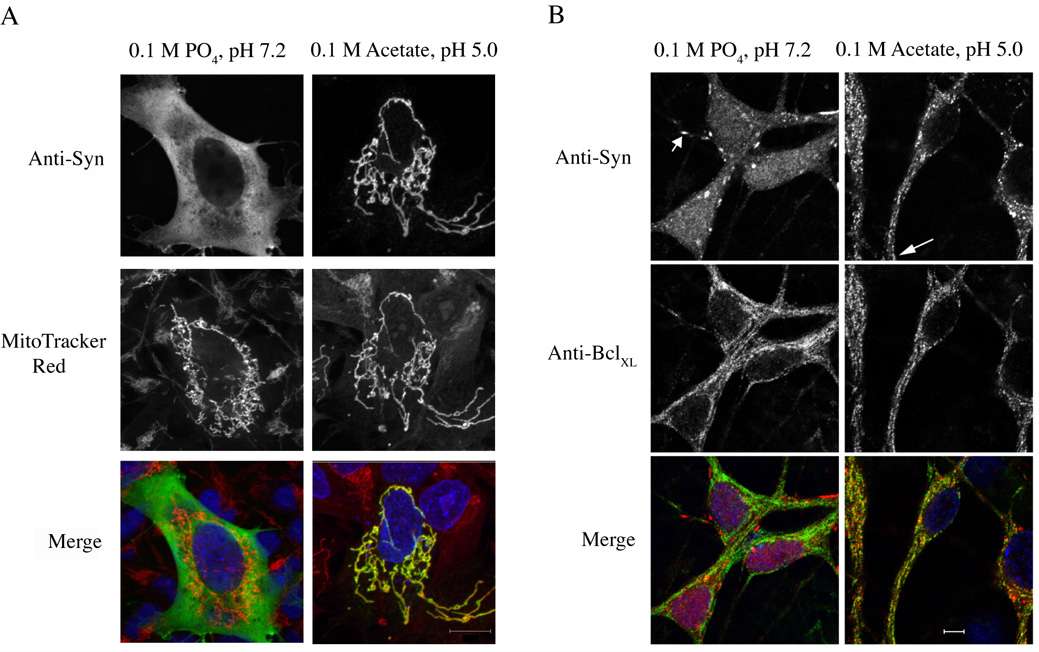
(A) Representative fluorescence micrographs of SK-N-SH cells transiently transfected with α-synuclein. Cells were fixed at pH 7.2 (left panels) or 5.0 (right panels). Synuclein is labeled with the antibody 202 and Alexa 488 donkey anti-mouse secondary antibodies. Mitochondria are labeled with MitoTracker Red. Merged images are shown at the bottom. Nuclei are stained with DAPI (blue). (B) Rat hippocampal neurons (4 div) were fixed at pH 7.2 (left panels) or pH 5.0 (right panels) and stained with antibodies to synuclein or to the mitochondrial protein BclXL. Short arrow indicates a synuclein-positive synaptic bouton; long arrow shows colocalization of synuclein and mitochondria in distal processes. Scale bar: (A) 10 µm; (B) 5 µm.