
Fig. 2.
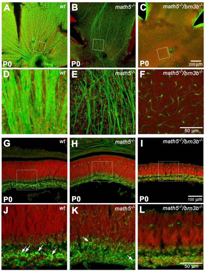
Projections of optical sections of flat-mounted and cross sectioned retinas at P0. Green: NF-L. Red: PI. Panels D, E, and F are enlarged from the boxed area of panels A, B, and C and rotated so that the optic discs or putative optics disc are at the bottom. Panels J, K, and L are enlarged from the boxed area of panels G, H, and I. A and D, well organized wildtype RGC axons can be seen at this stage. B and E, the number of NF-L-positive axons is drastically reduced. However, a significant amount of unorganized ones can be readily detected. C and F, scattered NF-L-positive cell bodies with short neurites can only be detected with high magnification. G and J, a cross sectioned wildtype retina showing newly-separated RGC layer. RGCs have distinct polygonal shapes of NF-L labeling, as indicated by the arrows. Amacrine cells above the emerging inner plexiform layer are also NF-L positive at this stage. The NF-L signal forms a thin circle, suggesting a unique cytoplasmic shape of amacrine cells. H and K, a Math5-deficient retina does not appear to be thinner than a wildtype control at this stage. However, besides a few possible RGCs (arrows), most of the cells in the INL have the shape of amacrine cells. I and L, the Math5/Brn3b-deficient retina is obviously thinner than the wildtype control. Cells exhibiting RGC shape cannot be found. Cell number in the RGC layer and NF-L positive cell number above the inner plexiform layer are severely reduced.