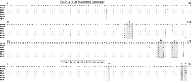
Figure 5. (a) Nucleotide and (b) amino-acid alignments of iguanine exon 3 (α-2 domain) sequences.
The lower-case letters and shaded boxes represent specific codon positions that show variation in amino acid residues between species. Species abbreviations are described in the Materials and methods section.

