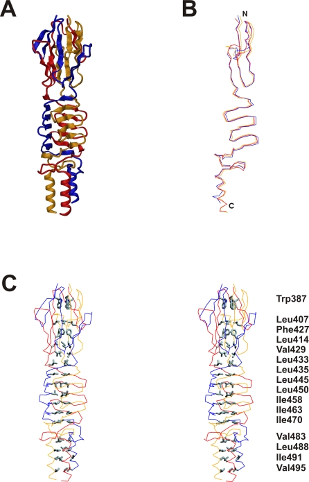
Figure 3. Quartenary structure of the BadA head domain.
(A) Structure of the entire BadA head fragment. The three independent protein chains are colored in yellow, red and blue. (B) Superposition of the three individual protein chains. A significant deviation is visible in particular at the N-terminal part of the structure. (C) Stereo representation of the hydrophobic core of the protein, which is built by 16 residues related by threefold symmetry.

