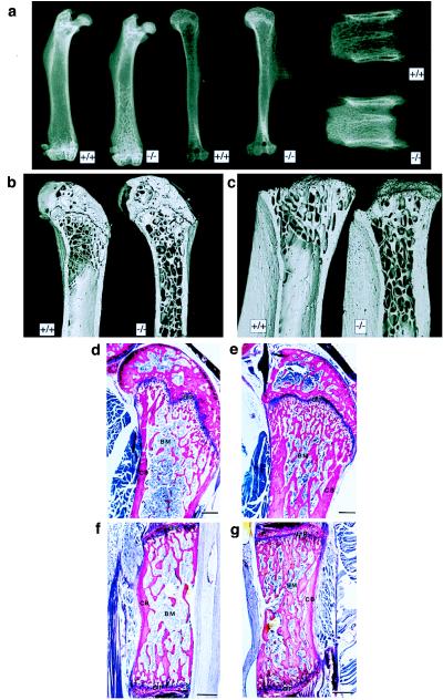
Figure 2.

Osteopetrosis in cathepsin-K deficient mice. (a) Radiographs of 16-week-old bones in cathepsin-K knockout (−/−) and control (+/+) mice. Left, femura. Center, humeri. Note extensive trabeculation of bone-marrow space (beginning at the distal end) in cathepsin-K-deficient mouse. Right, lumbar vertebrae (note the very dense and irregular distribution of spongiosa in the cathepsin-K-deficient mouse. (b and c) SEM images of 3-month-old proximal femura (b, with retained epiphyses) and tibiae (c, with epiphyses removed). Note the retention of cancellous bone in the shafts of cathepsin-K-deficient (−/−) bones. Field widths: b = 7.69 mm; c = 5.84 mm (d and e). Histological sections of the meta- and epiphysis of control (d) and cathepsin-K-deficient (e) femura embedded in methyl methacrylate and stained with McNeil’s tetrachrome. Note the unresorbed primary spongiosa in e. (f and g) Histological sections (sagittal plane) through the ventral half of thoracic vertebrae derived from control (f) and cathepsin-K-deficient (g) mice. BM, bone marrow; CB, cortical bone; GP, growth plate. [Bars = 400 μm (d–g).]
