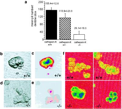
Figure 4.

Cathepsin-K-deficient osteoclasts with impaired bone resorption. Fresh osteoclasts were isolated and seeded as a suspension onto dentine sections, and (a) the total number of resorption pits therein was then determined. Cathepsin-K-deficient (−/−) osteoclasts formed significantly fewer resorption pits than did control (+/+) or heterozygote (+/−) ones. (Two-sample t test for +/+ vs. −/−: P = 0.0001; for ± vs. −/−: P = 0.001). (b and d) Images of resorption pits produced in the confocal reflection light microscope (maximum brightness; ref. 29). On dentine slices seeded with control osteoclasts (b), the pits are deeper and more sharply defined than on those seeded with cathepsin-K-deficient (−/−) osteoclasts (d). (c and e) Topographic-map images of resorption pits. Each band of color (pink, superficial; blue, deep) represents 1 μm in the vertical direction. The control pit (c), with characteristically steep sides, has a maximum depth of approximately 13 μm, whereas one of similar area created by a cathepsin-K-deficient osteoclast (e) is only about 3 μm deep. Field widths of b–e are 143 μm (f) Resorption pits analyzed by 15-kV digital backscattered electron imaging, pseudocolor coded to scale the amount of residual matrix. In cathepsin-K −/− pits, a greater thickness (more colors) of unprocessed residual matrix was found. Field width of each part = 128 μm.
