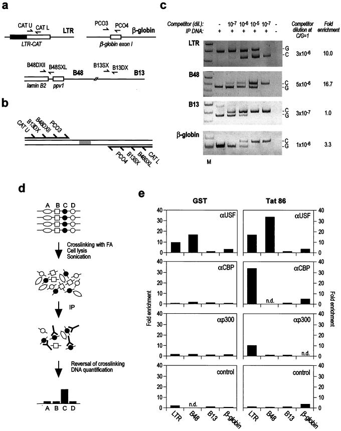
Figure 4.
Recruitment of p300 and CBP to the LTR upon Tat-mediated transcriptional activation in vivo. (a) Human chromosomal regions analyzed by quantitative chromatin immunoprecipitation. LTR-CAT, β-globin gene exon I, and lamin B2 gene B13 and B48 DNA segments were studied. For each of these regions, two primers were selected (small, converging arrows). The boxes schematically indicate the location of relevant genomic elements (LTR-CAT cassette, β-globin exon I, lamin B2 gene 3′ end, and ppv1 gene) with respect to primer localization. (b) Multicompetitor DNA for competitive PCR. The multicompetitor DNA fragment contains all primer recognition sites arranged to generate PCR products of size different from but comparable to those obtained from amplification of genomic DNA. (c) Quantification of the sample obtained from GST-treated HL3T1 cells immunoprecipitated with anti-USF antibody (e Top Left). Quantification of immunoprecipitated DNA was obtained by mixing a fixed amount of immunoprecipitated DNA with the indicated scalar amounts of competitor DNA, followed by PCR amplification with each primer pair. DNA quantification was obtained from the ratio between the amplification products for genomic (G) and competitor (C) DNAs. M, molecular weight markers. (d) Flow chart of the quantitative chromatin immunoprecipitation assay. A, B, C, and D indicate four genomic DNA segments that directly or indirectly are crosslinked to different proteins in vivo by treatment with formaldehyde (FA). When immunoprecipitation (IP) of sonicated chromatin is performed with an antibody reacting with the protein crosslinked to C, the immunoprecipitate will be enriched for this DNA segment. (e) Results of quantitative chromatin immunoprecipitations of the four analyzed regions after treatment of HL3T1 cells with GST (Left) or GST-Tat 86 (Right) and in the presence of chloroquine, using the indicated antibodies (control, antibody against the HA epitope). Results are expressed as fold enrichment with respect to B13 region. Antibody against USF immunoprecipitates crosslinked B48 and LTR-CAT regions but not B13 and β-globin; the effect is augmented by Tat treatment. Antibodies against CBP and p300 immunoprecipitate only crosslinked LTR-CAT DNA after Tat treatment. Control antibody failed to immunoprecipitate any of the four DNA regions after GST as well as Tat treatment. The graph reports the results obtained in a representative experiment. At least three independent experiments have been performed for each antibody and each DNA region, and consistent results were obtained. n.d., not done.

