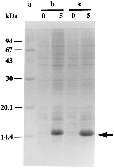
Figure 3.
Expression of streptavidin mutants Stv-A23D27 and Stv-A23E27. Lane a, molecular mass standard proteins; b, BL21(DE3)(pLysE)(pTSA-A23D27); and c, BL21(DE3)(pLysE)(pTSA-A23E27). Total cell protein at time 0 (at the time of induction; 133 μl of culture) and 5 hr after induction (67 μl of culture) was analyzed by SDS/PAGE. Proteins were stained with Coomassie Brilliant Blue. An arrow indicates the position where the streptavidin mutants migrate.

