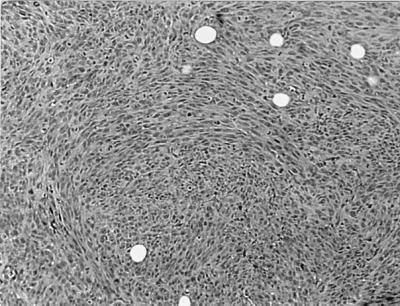
Figure 4.

Histology of tumor in MRG1–1-injected nude mice. Photomicrograph of tumor removed from mice injected with MRG1–1 cells, and stained with hematoxylin and eosin. Histologic pattern is sarcomatoid, consistent with the connective tissue origin of the parental cells. (Magnification: ×100.)
