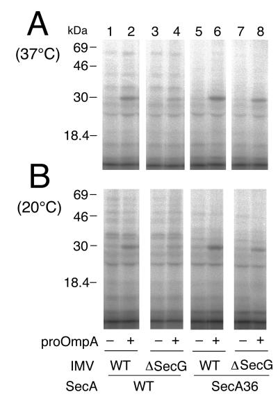
Figure 3.
Insertion of SecA+ and SecA36 into secG+ IMV vs. ΔsecG IMVs. 125I-SecA (100 μg/ml and 1.33 × 10⁵ cpm/μg; lanes 1–4) and 125I-SecA36 (100 μg/ml and 2 × 10⁵ cpm/μg; lanes 5–8) were subjected to incubation with urea-treated IMV (100 μg of protein per ml) from either the wild-type cells (GN67; lanes 1, 2, 5, and 6) or the ΔsecG∷kan cells (GN68; lanes 3, 4, 7, and 8), in the presence of ATP/ATP regeneration system and SecB, and in the presence (lanes 2, 4, 6, and 8) or absence (lanes 1, 3, 5, and 7) of proOmpA. After incubation at 37°C (A) or 20°C (B) for 15 min, samples were treated with trypsin. 125I-labeled protein fragments were separated by SDS/PAGE and visualized by a phosphorimager. The positions of molecular-mass markers are indicated on the left.

