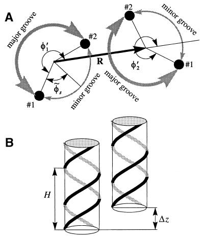
Figure 2.
An azimuthal (A) and axial (B) alignment of two neighboring B-DNA molecules. The sketch A is constructed as follows. We select the plane z = 0 at an arbitrary height. We then project the positions of two phosphates on molecule 1, each belonging to a different strand and nearest to the z = 0 plane. These projections are shown by the small, filled circles numbered as #1 and #2. We repeat the same projection procedure for molecule 2. We define the phosphate #1 on each molecule as the phosphate located at the major-to-minor groove crossover upon the counter clockwise rotation around the corresponding molecular axis. The angles φ′1 and φ′2 between the phosphates #1 and the direction of the vector R, defined as shown, give the azimuthal orientation of each molecule. The continuous helical lines in B are drawn through the centers of phosphate groups along the two strands of each molecule. H is the helical pitch, Δz is the axial shift needed to superimpose these helical lines after the lateral translation merging the axes of the molecules. The value of Δz characterizes the axial alignment.

