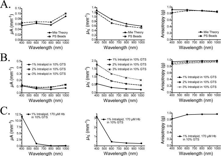
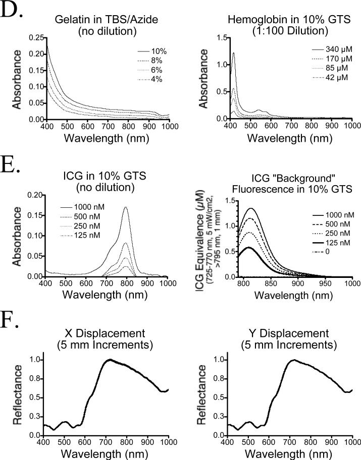
Figure 2. Optical Characterization of Phantom Components.
A) Validation of the employed inverse adding-doubling method by comparing measurements using polystyrene (PS) beads of defined optical properties (PS Beads) to Mie scattering theory (Mie Theory). Shown are μA (left), μS’ (middle) and anisotropy (right).
B) Optical properties of various intralipid concentrations as measured using an integrating sphere and the inverse adding-doubling method. Shown are μA (left), μS’ (middle) and anisotropy (right).
C) Optical properties of a typical phantom mixture compose of 1% intralipid and 170 μM Hb in 10% GTS. This composition most closely matches scattering human tissue with an 8% blood volume. Shown are μA (left), μS’ (middle) and anisotropy (right).
D) Absorbance of various gelatin concentrations in TBS/azide (left), and Hb in 10% GTS (right; diluted 1:100 in TBS/azide prior to measurement).
E) Absorbance (left) and “background” fluorescence (ICG Equivalence(725−775 nm, 5 mW/cm2, >795 nm, 1 mm), described in text; right) of ICG in 10% GTS.
F) Homogeneity of a 1% intralipid/170 μM Hb phantom in 10% GTS measured in 5 mm increments in the X (left) and Y (directions), over a total distance of 10 cm. Shown are the mean ± SEM of the 20 reflectance spectra at each wavelength from 400−1000 nm.


