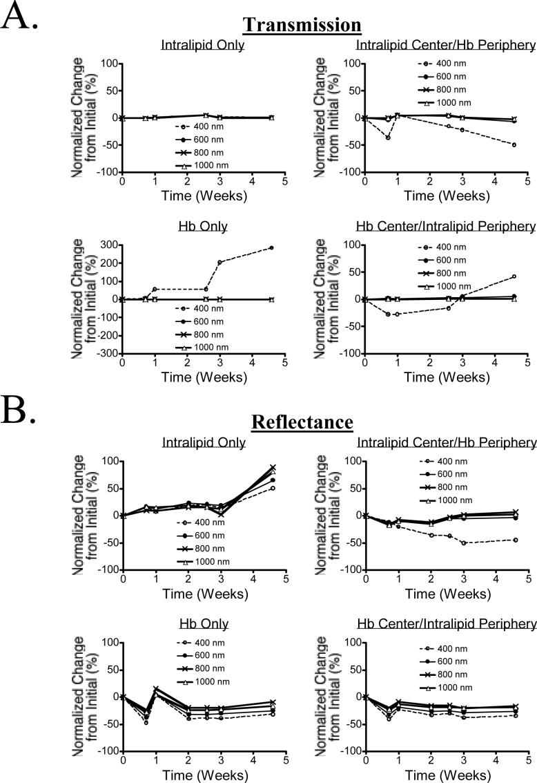
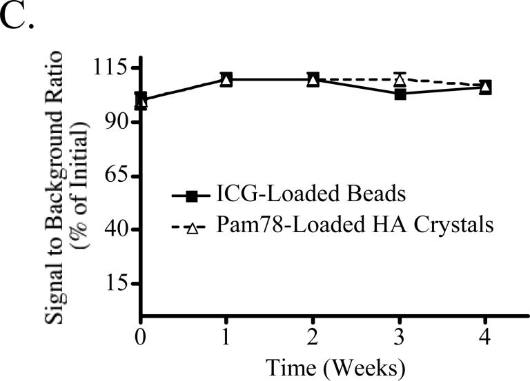
Figure 3. Long-Term Stability of Tissue-Like Phantoms and their NIR Fluorescent Inclusions.
A) Transmission over a 4-week interval for 1% Intralipid in 10% GTS (top left), 1% intralipid in a central location with Hb around the periphery, both in 10% GTS (top right), 170 μM Hb in 10% GTS (bottom left) and 170 μM Hb in a central location with 1% intralipid around the periphery, both in 10% GTS (bottom right).
B) Reflectance over a 4-week interval for the samples described in (A).
C) Stability of NIR fluorescent inclusions. NIR fluorescent beads and HA inclusions, each with a 1 μM ICG Equivalence(725−775 nm, 5 mW/cm2, >795 nm, 1 mm), were placed randomly throughout a phantom (1% intralipid and 170 μM Hb in 10% GTS) at a depth of 5 mm, and measurements were taken weekly. Between measurements, the phantom was sealed tightly in a storage container at 4°C. On the ordinate is signal to background ratio (SBR) expressed as mean ± SEM.


