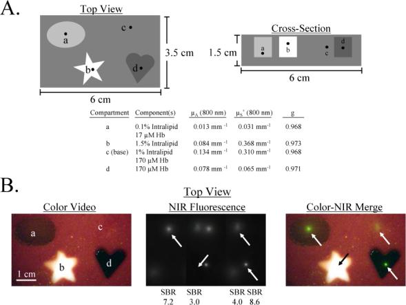
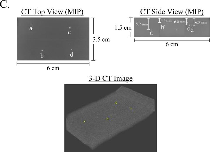
Figure 6. Complex Phantom for the Calibration and Assessment of a NIR Fluorescence Imaging System, and Independent Confirmation by Micro-Computed Tomography.


A) Schematic design of a complex phantom containing four compartments with optical properties shown in the table. Each compartment contained a 1 mm diameter bead of 880 nM ICG Equivalence(725−775 nm, 5 mW/cm2, >795 nm, 1 mm) and 88.8 mM Conray placed at the desired depth.
B) Simultaneous color video/ NIR fluorescence imaging (top view) of the phantom using a NIR excitation fluence rate of 5 mW/cm2 and 100 msec exposure time. Shown are color video (left), NIR fluorescence (middle), and a pseudocolored (lime green) merge of the two (right). All pixel values were within the linear range of the NIR camera. Below each NIR fluorescent bead (arrows) is shown its SBR.
C) Confirmation of precise position of NIR fluorescent inclusions using micro-computed tomography. Shown are the maximum intensity projections (MIP) for top view and side view, along with a 3-D rendering of the phantom with beads in yellow.
