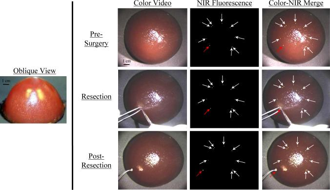
Figure 7. Training in Image-Guided Surgery using NIR Fluorescent Phantoms.
Nine 1 mm beads (each approximating a collection of 8 × 10⁵ cells; arrows) with a 1 μM ICG Equivalence(725−775 nm, 5 mW/cm2, >795 nm, 1 mm) were placed concentrically, and 0.5 cm below the surface, of a breast-shaped phantom (see oblique view). The phantom composition was 10% gelatin, 1% intralipid, and 17 μM hemoglobin, corresponding to μA = 0.056 mm−1, μS’ = 0.310 mm−1 and g = 0.968 at 800 nm. Shown is NIR fluorescence image-guided resection of one (red arrow) of the otherwise invisible beads using a scalpel and forceps. NIR fluorescence images have identical exposure times (100 msec) and normalizations.

