
Figure 2.
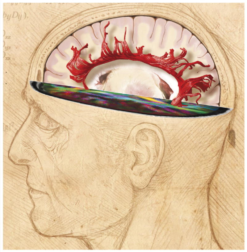
Neuroimaging reveals changes in white matter structure in the human brain. White matter (white) comprises half of the human brain and consists of bundles of myelinated axons connecting neurons in different brain regions. Gray matter (pink) is composed of neuronal cell bodies and dendrites concentrated in the outer layers of the cortex. Microstructural changes in white matter can be revealed by specialized MRI brain imaging techniques such as diffusion tensor imaging (DTI). This method analyzes the fractional anisotropy (FA) of proton diffusion in tissue, which is more restricted in white matter than in gray matter. The anisotropy increases with increased myelination, fiber diameter and axon compaction. The degree of anisotropy is represented on a pseudo color scale as shown in the human brain scan in the horizontal plane of the image above, where the major white matter tracts are revealed against a black background of low fractional anisotropy [107]. These data can be used to calculate the probable anatomy of white matter fiber bundles in living brain, a process called tractography. An example from human brain imaging is shown above as red filamentous bundles radiating out from the corpus callosum. Fiber orientation is calculated from the eigenvectors defining proton diffusion in three dimensions in each voxel. Using algorithms, the principal eigenvalue vector is connected to the next voxel to trace the fiber structure and orientation in white matter tracts [108]. Changes in white matter structure are seen by DTI in association with many neuropsychiatric disorders, cognitive function and during learning. FA image courtesy of Carlo Pierpaoli, NICHD, NIH, and DTI tractography, courtesy of Derek K. Jones, School of Psychiatry, Cardiff University. Illustration by Lydia Kibiuk, Medical Arts, NIH.