
Figure 3.
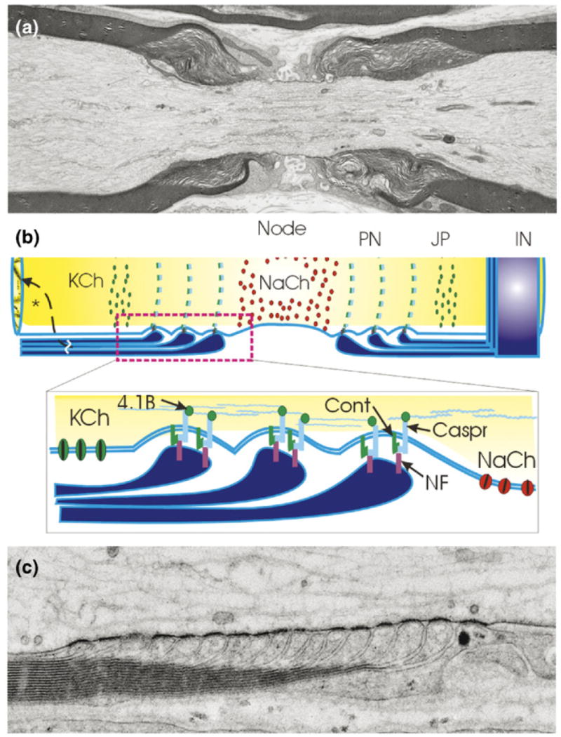
Myelin speeds impulse conduction velocity through nerve fibers by fundamentally changing the way impulses are propagated. Rather than a continuous wave of depolarization, the nerve impulse is generated by sodium and potassium currents at isolated points on the axon called nodes of Ranvier, and each node acts as a repeater. (a) An electron micrograph of a node of Ranvier in long section from spinal dorsal root nerve of rat showing the node of Ranvier flanked by intermodal segments insulated by layers of compact myelin. Each layer of myelin terminates in a series of loops adjacent to the node of Ranvier (the paranodal loops shown in [b] and [c]). (b) Three axonal domains are defined by axon interactions with myelinating glia: the Na+ channel-enriched node of Ranvier, the adjacent paranode (PN) where the loops of myelin adhere to the axon through cell-adhesion molecules linked to the axon cytoskeleton, the juxtaparanodal region (JP) which contains delayed rectifier K+ channels and the internode (IN) sealed by compacted layers of myelin membrane to restrict transmembrane ion currents to the nodal region. These domains are formed and maintained by adhesive interactions and soluble signals from myelinating glia. KCh = K+ channel; NaCh = Na+ channel; Cont = contactin; Caspr = contactin-associated protein; NF = neurofascin 155, 4.1B protein. (c) High-magnification electron micrograph of paranodal loops in a node of Ranvier from mouse spinal root nerve preserved by high-pressure freezing. Note the dense adhesive junctions between each paranodal loop and the axon. (a,c) Courtesy of Gina Sosinsky, Thomas Deerinck, Ying Jones and Mark Ellisman, UCSD, National Center for Microscopy and Imaging Research, San Diego. (b) Modified from Fields and Stevens-Graham [103].