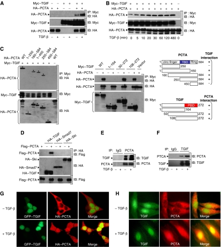
Figure 1.
PCTA interacts with TGIF. (A, B) 293 cells were transfected with Myc-TGIF in the absence or presence of HA-PCTA and treated with or without TGF-β for 1 h (A) or various times (B). Cell lysates were subjected to anti-Myc immunoprecipitation (IP) followed by immunoblotting (IB) with anti-HA. In these and all the following experiments, the expression of proteins under investigation was determined by direct immunoblotting. (C) 293 cells were transfected with Myc-TGIF and various HA-PCTA mutants (left) or HA-PCTA and various Myc-TGIF mutants (middle). Cell lysates were immunoprecipitated with anti-Myc and blotted with anti-HA. The TGIF-binding domain of PCTA (TBD, blue) and the PCTA-binding domain of TGIF (PBD, red) are indicated (right). (D) 293 cells were transfected with the indicated combinations of HA-TGIF, HA-Smad7, HA-Ski, and Flag-PCTA. Cell lysates were subjected to anti-HA immunoprecipitation followed by immunoblotting with anti-Flag. (E, F) 293 cells were treated with or without TGF-β for 1 h and cell lysates were immunoprecipitated with either rabbit IgG or anti-PCTA (E), and mouse IgG or anti-TGIF (F). TGIF-bound to PCTA (E) and PCTA-bound to TGIF (F) were detected by immunoblotting with anti-TGIF and anti-PCTA, respectively. (G) 293 cells were transfected with HA-PCTA and GFP–TGIF and treated with or without TGF-β for 1 h. Then, cells were immunostained with anti-HA and the localization of PCTA (red) or TGIF (green) was analysed by a fluorescence microscope. (H) Mv1Lu cells were treated with or without TGF-β for 1 h, immunostained with anti-PCTA and anti-TGIF, and the localization of PCTA (red) or TGIF (green) was visualized by a fluorescence microscope.

