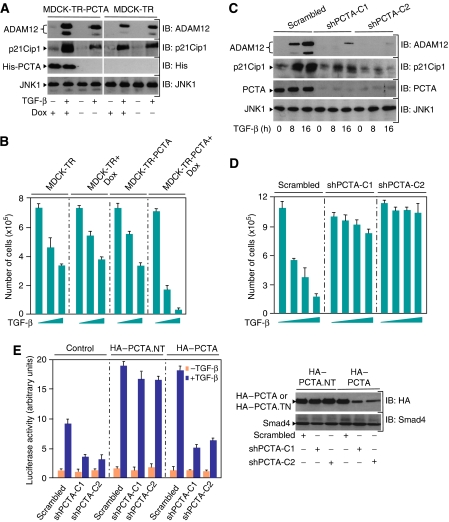
Figure 2.
PCTA contributes to TGF-β signalling. MDCK-TR or MDCK-TR-PCTA cells were treated with doxycyclin (Dox) for 24 h and then with or without 2 ng/ml TGF-β for 16 h (A) or with increasing amounts of TGF-β for 72 h (B). (A) Cell lysates were analysed by immunoblotting using antibodies against PCTA, p21Cip1, ADAM12 and JNK1 as a loading control. (B) Cells were counted and the results were expressed as mean±s.d. of triplicate from a representative experiment performed at least three times. (C) MDCK-shPCTA (clones 1 and 2) or MDCK-Scrambled cells were treated with or without 2 ng/ml TGF-β for 8 or 16 h (C) or with increasing amounts of TGF-β for 72 h (D). (C) Cell lysates were analysed by immunoblotting using antibodies against PCTA, p21Cip1, ADAM12, and JNK1. (D) Cells were counted and the results were expressed as mean±s.d. of triplicate from a representative experiment performed at least three times. (E) MDCK-shPCTA (clones 1 and 2) or MDCK-Scrambled cells were transfected with ARE3-Lux together with FAST1 and either HA-PCTA or the non-targetable form HA-PCTA.NT. Cells were treated with or without TGF-β for 16 h and analysed for luciferase activity (left). Cell extracts were analysed by immunoblotting with anti-HA or anti-Smad4 as a loading control (right).

