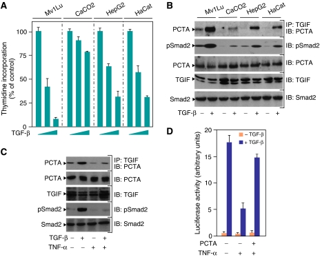
Figure 4.
Physiological relevance of the PCTA function in TGF-β signalling. (A) Mv1Lu, CaCO2, HepG2, and HaCat cells were treated with increasing amounts of TGF-β for 48 h and the rate of cell proliferation was determined by the thymidine incorporation method. (B) Mv1Lu, CaCO2, HepG2, and HaCat cells were treated with or without TGF-β for 1 h. PCTA bound to TGIF was detected by blotting anti-TGIF immunoprecipitates with anti-PCTA. To detect Smad2 phosphorylation, cell lysates were blotted with anti-pSmad2. (C) MEFs were treated with TNF-α for 8 h prior to treatment with TGF-β for 1 h. Cell extracts were immunoprecipitated with anti-TGIF followed by blotting with anti-PCTA. To detect Smad2 phosphorylation, cell lysates were blotted with anti-pSmad2. (D) MEFs were transfected with ARE3-Lux together with FAST1 in either the presence or absence of PCTA. After 24 h, cells were treated with the indicated combinations of TGF-β and TNF-α for 16 h and were analysed for luciferase activity.

