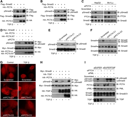
Figure 5.
PCTA promotes TGF-β-mediated phosphorylation of Smad2. (A, B) 293 cells were transfected with Flag-Smad2 (A) or Flag-Smad3 (B) in either the presence or absence of HA-PCTA and treated with or without TGF-β for 1 h. The phosphorylation of Smad2 (A) or Smad3 (B) was assessed by blotting anti-Flag immunoprecipitates with anti-pSmad2 or anti-pSmad3, respectively. (C) HepG2 or Mv1Lu cells were transfected with either Scrambled or PCTA siRNA and 48 h later, they were treated with or without TGF-β for 1 h. The phosphorylation of Smad2 was assessed by immunoblotting with anti-pSmad2. (D) 293 cells were transfected with the indicated constructs, treated with or without TGF-β for 1 h and the phosphorylation of Smad2 was assessed by blotting anti-Myc immunoprecipitates with anti-pSmad2. (E–G) MDCK-shPCTA (clones 1 and 2) or MDCK-Scrambled cells were treated with or without TGF-β for 1 h. (E) The phosphorylation of Smad2 was assessed by immunoblotting with anti-pSmad2. (F) The association of Smad2 with Smad4 was analysed by blotting anti-Smad2 immunoprecipitates with anti-Smad4. (G) The localization of Smad2 was revealed by immunofluorescence with anti-Smad2. (H) 293 cells were transfected with the indicated combinations of Myc-Smad2, HA-TGIF, and HA-PCTA and treated with or without TGF-β for 1 h. The phosphorylation of Smad2 was assessed by blotting anti-Myc immunoprecipitates with anti-pSmad2. (I) MDCK-pSUPER or MDCK-pSUPERTGIF cells were transfected with the indicated siRNA and treated with or without TGF-β for 1 h. The phosphorylation of Smad2 and Smad3 was assessed by immunoblotting with anti-pSmad2 and anti-pSmad3.

