Figure 6.
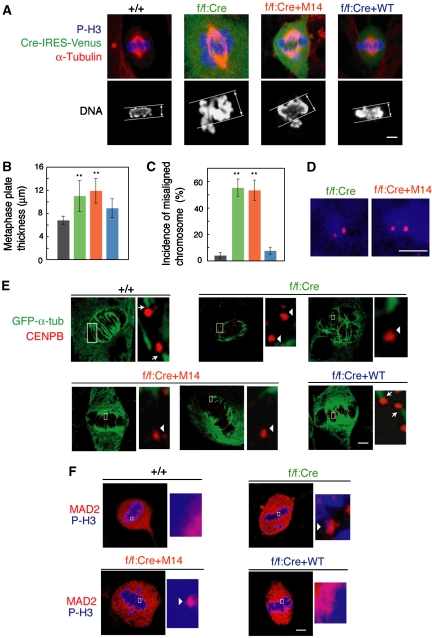
The Myb–Clafi complex is required for the function of the mitotic spindle. (A) Thicker metaphase plates in M14-reexpressing cells and the B-Myb-deficient cells. The indicated MEFs were immunostained with anti-α-tubulin (red). DNA was stained with TOTO-3 (blue). Cells expressing Cre recombinase together with Venus were immunostained with anti-GFP (green). Signals were visualized with appropriate secondary antibodies using confocal microscopy. Scale bars: 5 μm. (B) Increased thickness of the metaphase plate in M14-reexpressing cells and the B-Myb-deficient cells. The thickness of the metaphase plate ±s.d. (n=30) is shown. **P<0.001. (C) Increase in misaligned chromosomes in M14-reexpressing cells and the B-Myb-deficient cells. The frequency of metaphase-like cells with misaligned chromosomes ±s.d. (n=100–120) is shown. (D) Misaligned chromosomes in M14-reexpressing cells and B-Myb-deficient cells were pairs of sister chromatids. Cells were stained with anti-CENPB (red) and anti-phospho-histone H3 antibody (blue). Misaligned chromosomes were not obviously detected in the wild-type control (+/+) cells. Scale bars: 5 μm. (E) Failure in attachment of kinetochores to microtubules in M14-reexpressing cells and B-Myb-deficient cells. The indicated cells were stained for CENPB (red) and α-tubulin (green), after cold treatment to depolymerize non-kinetochore microtubules. The right panels show higher magnification images of the centromeres delineated by the white box. The arrows indicate the centromeres that are normally connected with α-tubulin, whereas the arrowheads indicate the centromeres that are not connected with α-tubulin. Scale bars: 5 μm. (F) Persistent localization of Mad2 on kinetochores of M14-reexpressing cells and B-Myb-deficient cells. Cells were stained with anti-Mad2 (red) and anti-phospho-histone H3 (blue). The right panels show higher magnification images of the kinetochores delineated by the white box. Mad2-positive kinetochores are indicated by the arrowheads. Scale bars: 5 μm.

