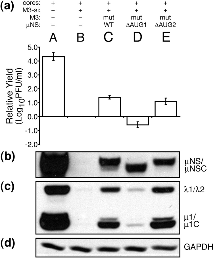
Fig. 8.
Expression of μNSC in the absence of μNS is unable to rescue viral growth. BSRT7 cells were transfected using Lipofectamine 2000. Lanes reflect different combinations of transfected materials as follows: (A) T1L cores alone; (B) T1L cores and M3-si01; (C) T1L cores, M3-si01, and mutRNA expressing wild-type μNS (and μNSC) as in Fig. 4; (D) T1L cores, M3-si01, and mutRNA expressing μNSC only (ΔAUG1); and (E) T1L cores, M3-si01, and mutRNA expressing full-length μNS only (ΔAUG2). Cells were harvested at 24 h p.i. for viral titers (a) and protein analyses (b–d) as described for Fig. 4. Titers were expressed as relative yields as described for Fig. 5. The blots are representative of the three replicate experiments.

