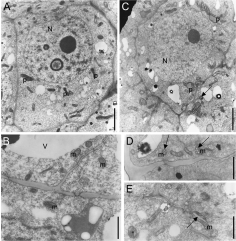
Fig. 1.
Altered ultrastructure of cells in shoot meristems of cytokinin-deficient tobacco plants. Transmission electron micrographs of cells from the peripheral zone of wild-type (A, B) and 35S:CKX1 transgenic shoot meristems (C–E) are presented at a similar magnification. N, Nucleus; m, mitochondria; p, proplastid; V, vacuole. Arrows in C, D, and E indicate the unusually large tubuli arising from the inner membrane of mitochondria, sectioned transversally (C, D) and longitudinally (E). Scale bars = 2 μm (A, C) and 1 μm (B, D, E).

