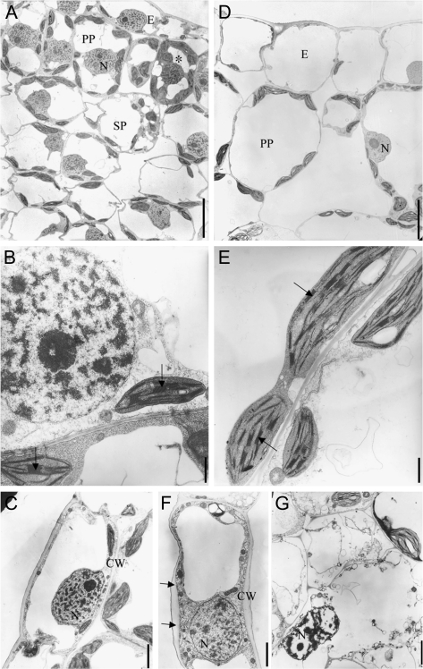
Fig. 2.
Altered cellular ultrastructure in developing leaves of cytokinin-deficient plants. Transmission electron micrographs of young leaves (leaf 3; see Fig. 3) from wild-type (A–C) and 35S:CKX1-expressing (D–G) tobacco plants. Cross-section of wild-type (A) and 35S:CKX1 (D) leaf. The asterisk indicates a cell in preparation for mitosis with dense cytoplasm and condensed chromatin. (B, E) Comparison of chloroplast structure (arrows indicate grana). (C, F) Comparison of adaxial epidermal cells (arrows point to irregular cell wall thickness). (G) Necrotic cell with pycnotic nuclei and disorganized plastid and membrane. CW, Cell wall; E, epidermis; N, nucleus; PP, palisade parenchyma; SP, spongy parenchyma. Scale bars = 10 μm (A, D), 2 μm (B, E), and 1 μm (C, F, G).

