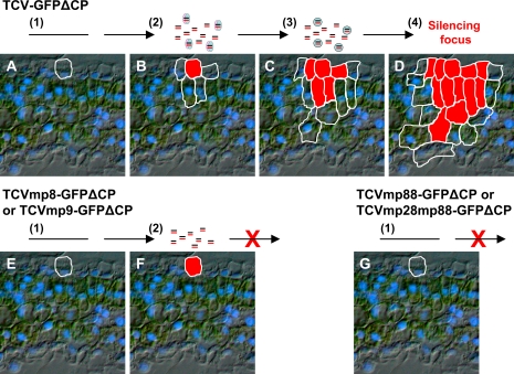
Fig. 6.
Schematic model for p8- or p9-mediated cell-to-cell spread of RNA silencing. (A–D) Induction and intercellular spread of RNA silencing induced by TCV–GFPΔCP. (E, F) RNA silencing in single-epidermal cells infected by TCVmp8–GFPΔCP or TCVmp9–GFPΔCP. (G) No RNA silencing induction by TCVmp88–GFPΔCP or TCVmp28mp88–GFPΔCP. The sequences of events are as follows: (1) viral RNA transcript enters an epidermal cell following mechanical inoculation (A, E, G); (2) viral RNA then replicates and triggers RNA silencing in the first invaded epidermal cell (B, F). At this stage, the primary silencing signals are produced. Without replication, these viruses cannot trigger RNA silencing (G). ‘=’ stands for mobile RNA silencing signal, ellipses for p8 or p9, and circles for host factors required for silencing cell-to-cell movement.

