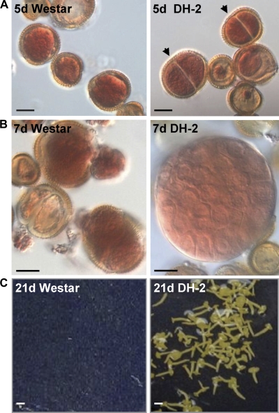
Fig. 1.
Microspore-derived embryo development in Brassica napus cv. Westar and the Westar-derived DH-2 line. (A) Acetocarmine-stained 5 d enlarged microspores. Arrowheads indicate divisions in the microspores of the DH-2 line. (B) Acetocarmine-stained 7 d enlarged microspores in Westar and a dividing pre-globular embryo in the DH-2 line. (C) Twenty-one day mid-maturation stage embryos in the DH-2 line; no embryos developed in this microspore culture plate of Westar. Black bars=10 μm, white bars=35 μm.

