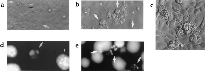
Figure 1.
(a) Light microscopic view (×600) of 24-hr coculture of wild-type hepatocytes and control fibroblasts. Arrows indicate normal hepatocytes. ∗ indicates fibroblast monolayer. (b) 24-hr coculture of wild-type hepatocytes with FasL-expressing fibroblasts (×600). Arrows point to dying hepatocytes. (c) Light microscopic view of blebbing hepatocytes with granular cytoplasm on the surface of monolayer of FasL-expressing fibroblasts (original magnification ×600, digitally enlarged). (d) DAPI (4′,6′-diamidino-2-phenylindole) staining illustrating hepatocyte nuclear condensation and (e) 24-hr coculture of wild-type hepatocytes with FasL-expressing fibroblasts (×320).

