Figure 5.
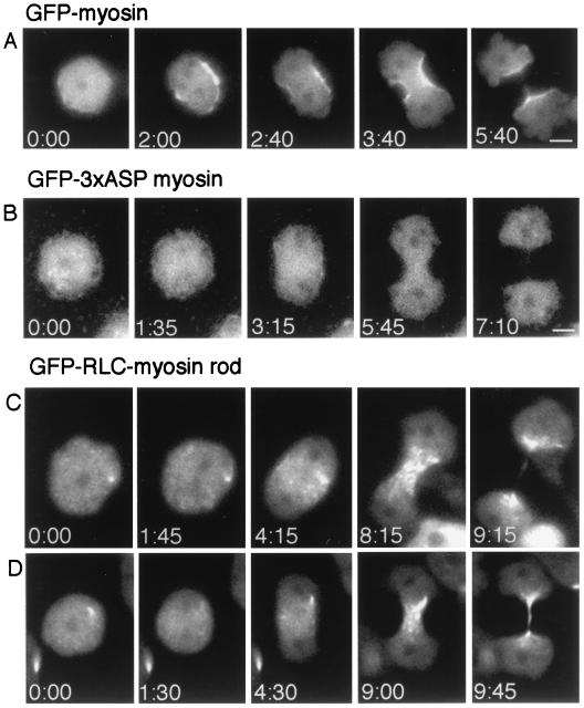
Myosin II-null cells expressing various GFP proteins undergoing division on a surface. GFP-myosin II and GFP-RLC-myosin rod, but not GFP-3xASP myosin II, are localized to the cleavage furrows. (A and B) Time-lapse fluorescence microscopy captures division of a cell expressing GFP-myosin II, which is localized to the cortex of the cleavage furrow (A), and division of a cell expressing GFP-3xASP-myosin II, which is diffuse throughout the cell during the adhesion-dependent, myosin II-independent cytokinesis B process (B) (5). (Bars, 5 μm.) (C and D) Symmetrical divisions by the myosin II-independent cytokinesis B process in which the GFP-RLC-myosin rod is distributed equally to daughter cells of similar sizes. GFP-RLC-myosin rod is localized to the cleavage furrow region, but differs from GFP-myosin II in that GFP-RLC-myosin rod is not cortical.

