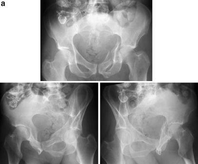
Fig. 3.
a An active 58 year-old male was struck by an automobile while bicycling. Anteroposterior (AP) and Judet radiographs revealed a left-sided comminuted both column acetabular fracture. No associated injuries were present and past history was significant only for previous aortic valve replacement. He was treated with ORIF via an ilioinguinal approach three days following the injury. EMG and SSEP monitoring were used during the procedure. b CT-scan images further delineate the fracture lines. c,d The patient was treated with ORIF via an ilioinguinal approach three days following the injury with EMG and SSEP used during the procedure. Intraoperative fluoroscopic images (c) illustrate an anatomic reduction and AP radiograph at 6 weeks (d) demonstrate maintenance of fixation. e Postoperative CT-scan images at 2 days following acetabular surgery demonstrating congruency and acceptable hardware placement. f AP and Judet views at 57 months following ORIF illustrate a healed acetabular fracture and an excellent radiographic result. The patient reported complete pain resolution and a return to participation in cross-country bicycling

