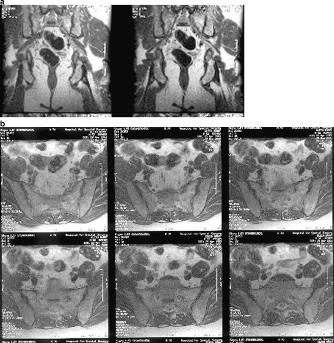
Fig 1.
MRI pelvis, non-contrast: (a) coronal and (b) axial images of the pelvis show a 2.8-cm intermediate focus of abnormal signal probably representing a mass associated with a thickened left S1 nerve as it courses out of the foramen. Sciatic nerve shows high signal extending through its distal course, but no distal thickening

