Figure 6.
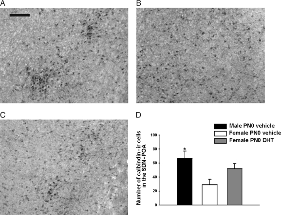
A–C, Representative photomicrographs of calbindin D28K-ir cells in adult mouse brains. These cells are observed in the boundary between the anterior preoptic area and the anterior hypothalamus in each experimental group. A, A male treated on PN0 with vehicle. B, An identically treated female. C, The calbindin-ir in a female that received DHT on PN0. Scale bar, 100 μm. D, The mean (±sem) numbers of calbindin-ir cells present in the mPOA. In this study we used brains from five control males, nine brains from vehicle-treated control females, and 10 females treated with DHT on PN0. At the time the animals were killed, all adults were gonadectomized and had been without hormone treatment for at least 2 wk. *, Significantly different from the vehicle-treated female group (P < 0.05).

