Figure 6.
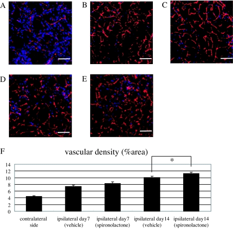
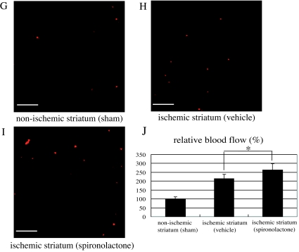
Effects of spironolactone on vascular regeneration and blood flow in the ischemic striatum after 20-min MCAo. A–E, Histological examination of the vasculature in the ischemic core stained with mouse PECAM-1 (red) and Neu-N (blue). Shown are representative photomicrographs in the nonischemic striatum (A) and the ischemic striatum of vehicle- (B and D) and spironolactone-treated (C and E) mice on d 7 (B and C) and 14 (D and E) after MCAo. F, Quantitative analysis of the relative area of PECAM-1 positivity (percent area) in the nonischemic striatum (n = 5) and ischemic striatum of vehicle- and spironolactone-treated mice (n = 9–10) on d 7 and 14 after MCAo. G–I, Representative photomicrographs of sections of the nonischemic striatum of a sham-operated mouse (G) and the ischemic core of the striatum in vehicle- (H) and spironolactone-treated (I) mice on d 14 after MCAo. J, Quantitative analysis of the relative blood flow in the nonischemic striatum of sham-operated mice (n = 5) and ischemic core of the striatum in vehicle- (n = 11) and spironolactone-treated mice (n = 11) on d 14 after MCAo. The relative blood flow in the ischemic striatum in vehicle- and spironolactone-treated mice was expressed as the ratio of number of fluorescent microspheres in the ischemic core to that in the nonischemic striatum of sham-operated mice (set as 100%). *, P < 0.05 spironolactone vs. vehicle. Scale bar, 100 μm (A–E, G–I); magnification, ×20.


