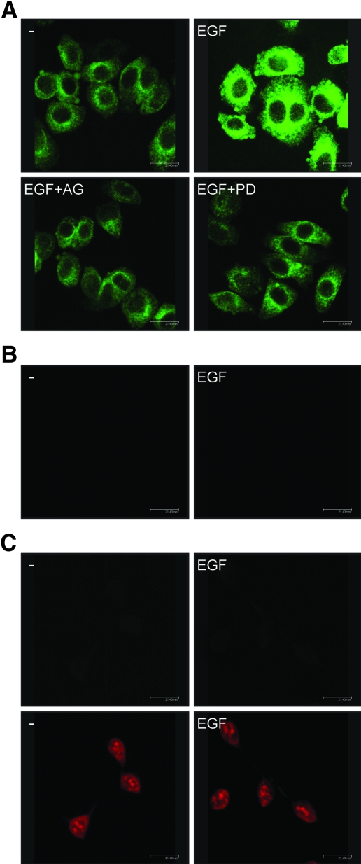
Figure 5.
GPR30 localization in SkBr3 cells. A, GPR30 evaluation by confocal microscopy in SkBr3 cells fixed, permeabilized, and stained with anti-GPR30 antibody. Cells were treated for 2 h with vehicle (−) or 50 ng/ml EGF alone and in combination with EGFR inhibitor tyrphostin AG, 10 μm MEK inhibitor PD, as indicated. B, SkBr3 cells were treated for 2 h with vehicle (−) or 50 ng/ml EGF and stained with GPR30 antibody, which was preneutralized with the antigen peptide. C, HEK-293 cells were treated for 2 h with vehicle (−) or 50 ng/ml EGF and stained with GPR30 antibody (upper panels) or propidium iodide (lower panels). The white bars denote 21,43 μm. Data are representative of three independent experiments.

