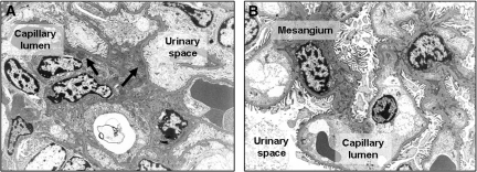
Figure 2.
Rapamycin started on the day of immunization markedly inhibits morphologic changes of GN. Representative electron microscopy pictures from day 21 are shown. (A) Ultrastructurally, active immunocomplex GN with mesangial widening, hypercellularity, partial podocyte foot process effacement, and abundant electron-dense immunocomplex deposits (black arrows) were detected in mice that received vehicle and were subjected to anti-GBM GN. (B) Electron microscopy of rapamycin-treated mouse kidneys revealed regular glomerular ultrastructure with narrow mesangia, regular podocyte foot processes, and inconspicuous endothelial cells. No immunocomplex deposits were detected. Magnification, ×5000.

