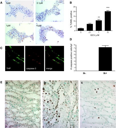
Figure 2.
Apoptosis studies. (A and B) In vitro studies. Representative fields from TUNEL assays on HEK-HSV1-tk stably transfected cells treated for 48 h with the doses of GCV indicated. (B) Quantitative analysis of percentage TUNEL positive. **P < 0.01 versus 0 μM; ***P < 0.001 versus 0 μM. (C) In vivo studies on GCV-treated kidneys co-stained for activated caspase-3 and THP. THP delineates TAL, and apoptosis was seen as nuclear staining in tubules that stain for THP. (D) Quantitative analysis of activated caspase-3. (E) TUNEL on kidney sections confirms the presence of apoptosis. (a) GCV-treated tk− kidneys had no evidence for TUNEL reactive cells. (b and c) GCV-treated tk+ kidneys show TUNEL-reactive cells in the tubular epithelia (b) as well as detached cells within tubular lumens (c; oil). Magnifications: ×630 in A and E, c; ×400 in C.

