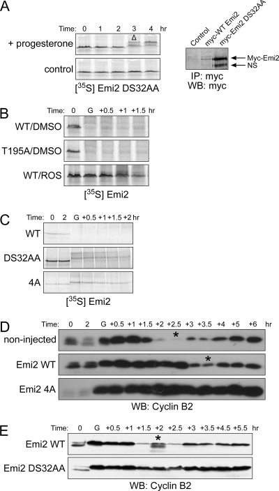
Figure 2.
Emi2 degradation is mediated through Cdc2 phosphorylation on 213/239/252/267 sites. (A) Left, 35S-labeled DS32AA Emi2 protein was injected into oocytes, and samples were processed as in Figure 1C. GVBD was monitored visually. Δ, GVBD. Right, oocytes were injected with Myc6-Emi2-3′-UTR mRNA (wild-type or DS32AA). Samples were processed same as in Figure 1A. NS, nonspecific band. (B) Oocytes were injected with 35S-labeled Emi2 (WT or T195A). One hour after injection, oocytes were treated with progesterone in the presence or absence of 300 μM roscovitine. At the indicated times, lysates were made and analyzed by SDS-PAGE and autoradiography. GVBD was monitored visually. G, GVBD. (C) Oocytes were injected with 35S-labeled Emi2 (wild-type, DS32AA or 4A). One hour after protein injection, oocytes were treated with progesterone and samples were processed as in Figure 1B. (D) Oocytes were injected with Flag-Emi2 mRNA appended with β-globin 3′-UTR (wild-type or 4A; 0.3 ng/oocyte). After overnight incubation, oocytes were treated with progesterone, and samples were processed as in Figure 1D. (E) Oocyte were injected with Myc6-Emi2-3′-UTR mRNA (0.3 ng/oocyte) and incubated for 1 h. before progesterone treatment. Samples were processed as in Figure 1D.

