Figure 2.
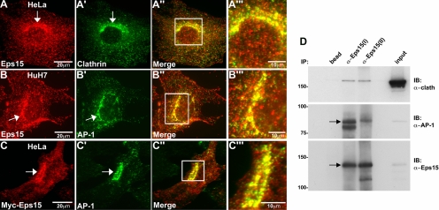
Eps15 colocalizes with clathrin and the adaptor protein complex AP-1 at the Golgi. (A–B‴) HeLa cells were immunostained with antibodies against Eps15 and clathrin (A–A‴), whereas HuH7 cells were immunostained with antibodies against Eps15 and AP-1 (B–B‴). (C–C‴) Additionally, HeLa cells expressing Myc-tagged Eps15 were immunostained for Myc and AP-1. Colocalization of Eps15 (A, B, and C) with clathrin (A′) or AP-1 (B′ and C′) at the Golgi region is marked by arrows. Higher magnification images (A‴, B‴, and C‴) of regions boxed in white in the respective merged images shown to the left (A″, B″, and C″) show substantial overlap (yellow) of Eps15 (red) with clathrin (A‴, green) or AP-1 (B‴ and C‴, green) at the perinuclear Golgi region. (D) Protein extracts of fractionated Golgi from rat liver were subjected to immunoprecipitation using two different anti-Eps15 antibodies [anti-Eps15(I) or anti-Eps15(II)] and subsequently analyzed by Western blot, probing with antibodies against clathrin, AP-1, and Eps15. The last lane (input) represents 5% of the total protein extract used for immunoprecipitation. A substantial amount of AP-1 as well as a modest amount of clathrin heavy chain coimmunoprecipitate with either of the anti-Eps15 antibodies. Scale bars, 20 μm (A, B, and C); 10 μm (A‴, B‴, and C‴).

