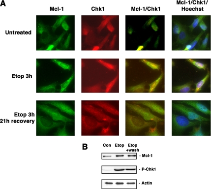
Figure 4.
Mcl-1 and Chk1 colocalize after DNA damage. (A) HeLa cells were untreated, treated with 15 μM etoposide for 3 h, or treated with etoposide for 3 h, followed by washing and allowing cells to recover for 21 h. Cells were stained using anti-Mcl-1 or anti-Chk1, or the figures were merged to show colocalization. Additional staining with Hoechst 33342 was used to visualize nuclei. (B) Cell extracts from the same cells used in A were used to immunoblot for the presence of Mcl-1, phospho-Chk1, or actin as a loading control.

