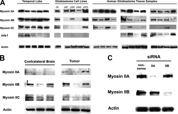
Figure 2.
Myosin II isoform expression in gliomas. (A) Immunoblot myosin II isoforms from glioma cell and human tissue lysates. β-Actin was used as a loading control. (B) Comparison of myosin II isoform expression in lysates from the rat glioma model generated by intracerebral injection of a PDGF-IRES-GFP–encoding retrovirus, compared with the uninjected contralateral brain. Immunoblot analysis was performed on excisions from three separate rat brains. (C). Immunoblot analysis of myosin IIA and myosin IIB siRNA-transfected U251 cells showing >95% reduction of each isoform. Nonsense siRNA oligos were used as a control.

