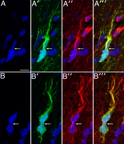
Figure 5.
Infiltrating human glioma cells undergo the same cell body and nuclear deformation seen in the PDGF-driven glioma model. Panels A and A′ show a GFP-expressing human glioma cell (green) infiltrating the surrounding, normal brain, with nuclear DAPI staining in A and GFP staining in A′. There is strong immunostaining for myosin IIA (red, A″), and these three images are merged in A‴. Bar, 10 μm. B and B′ correspond to DAPI and GFP staining, respectively, of another infiltrating GFP-expressing human glioma cell. This cell demonstrates strong staining for myosin IIB (red, B″). In both sets of micrographs the white arrows point to focal deformation of the cell bodies.

