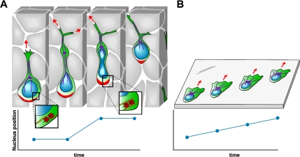
Figure 8.
Model of how a glioma cell migrates through brain tissue. (A) Starting with the left-most image, the infiltrating glioma cell is depicted as having a prominent leading process that often branches at its distal end. A dilatation forms in the leading process (black arrow). Evidence form neural progenitors show that the centrosome and associated microtubules (purple) move forward into the dilatation (Tsai et al., 2007). The mechanical constraints generated from the small intercellular spaces impede forward movement of the nucleus and cell body until contraction of actomyosin II at the rear of the cell (red) provide the necessary force to squeeze the nucleus through a narrowing in the extracellular space. The graph of nuclear position over time illustrates the resulting saltatory movement. (B). Glioma cells crawling on a two-dimensional surface move in a different manner that more closely resembles the migration of fibroblasts. Migration is associated with formation of broad lamellipodium, the nucleus remains undistorted, and its forward movement is continuous and unimpeded, as illustrated by the linear plot of nuclear position over time. Although myosin II is involved in maintaining cell polarity and shape, its activity is not required for cell motility in this barrier-free environment.

