Figure 1.

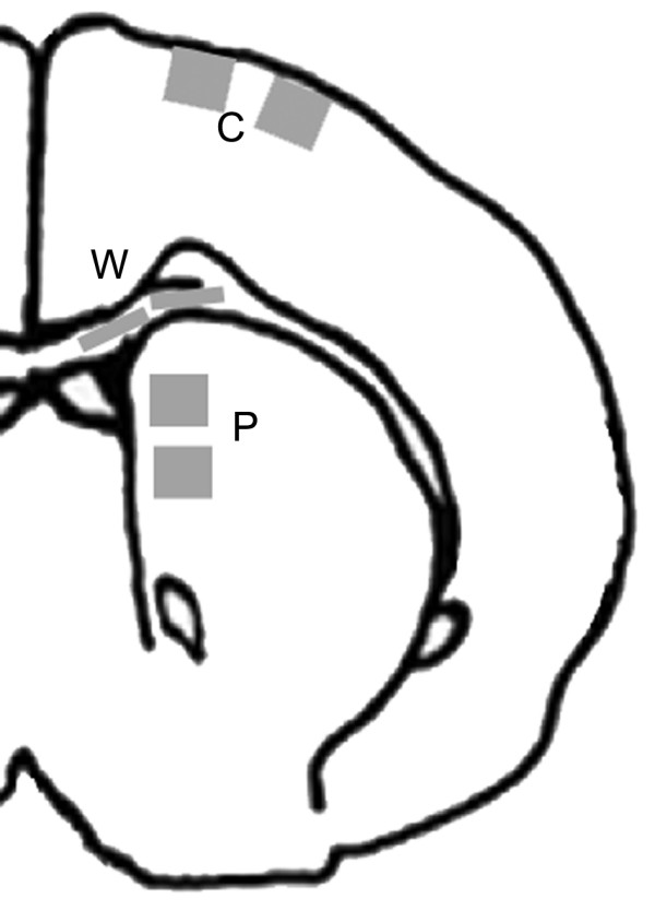
Schematic diagram of rat brain showing shaded areas in which cerebral cortex (C), periventricular white matter (W), and medial caudatoputamen (P) were photographed for image analysis of histochemical staining and immunolabeling.

Schematic diagram of rat brain showing shaded areas in which cerebral cortex (C), periventricular white matter (W), and medial caudatoputamen (P) were photographed for image analysis of histochemical staining and immunolabeling.