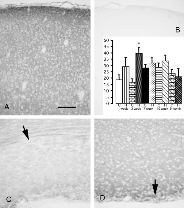
Figure 3.
Photomicrographs showing immunolabeling for neurocan of rat cerebral cortex at 3 weeks of age. The cerebral cortex of a control brain (A) shows a diffuse extracellular pattern, most prominent in the glia limitans. Hydrocephalic brains appear the same (not shown). Omission of the primary antibody is associated with no labeling (B). Normal white matter (C, arrow) is less intensely labeled. White matter in hydrocephalic brains (D, arrow) is atrophic and the residual tissue is more intensely labeled. Scale bar = 100 μm. Bar graph shows densitometric analysis of immunolabeling (arbitrary units) in white matter of control (C) and hydrocephalic (H) rats. The hydrocephalus-related increase is significant only in the 3-week rats (*p = 0.0007 ANOVA, Bonferroni-Dunn; all groups n = 4). No significant changes were observed in any other location.

