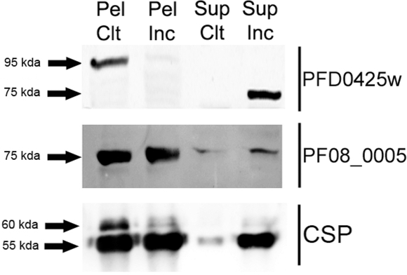
Figure 3. Detection of PFD0425w (SIAP-1) and PF08_0005 (SIAP-2) proteins in P. falciparum sporozoites.
Western blots of pellets (Pel) or supernatants (Sup) from control salivary gland sporozoites (Ctl) or those incubated for 2 hours at 37°C (Inc), probed with both specific antisera or anti-PfCSP monoclonal antibody (lower panel). Arrows indicate the positions to which size markers had migrated.

