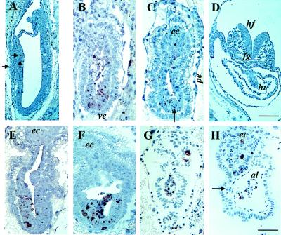
Figure 3.

Histological analysis of E7.5 and E8.5 embryos. Paraffin sections (4 μm) of embryos were subjected to TUNEL reaction as described (15). Embryos in A and F were obtained from inbred strains. (A) E7.5 wild-type embryo. Solid arrows point to apoptotic bodies present in the anterior embryonic ectoderm. The section in A is adjacent to the sections presented in Fig. 4 A and C. (B and C) E7.5 mutant littermates. The section in C is adjacent to the sections presented in Fig. 4 B and D. The arrow points to a presumptive primitive streak. (D) E8.5 wild-type embryo. (E and F) E8.5 mutant embryos, littermates of the embryo shown in D. (G and H) E7.5 and E8.5 mutant embryos, respectively, from the outbred strain Black Swiss. The solid arrow in H points to a presumptive blood island. [Bar = 100 μm in D (also applies to A) and 50 μm in H (also applies to B, C, E, F, and G).] Abbreviation used in Figs. 3 and 4 are as follows: ab, allantoic bud; al, allantois; amn, amnion; ec, ectoplacental cone; ee, embryonic ectoderm; ems, embryonic mesoderm; fg, foregut; hf, headfold; ht, heart; nch, notochordal plate; pe, parietal endoderm; ve, visceral endoderm.
