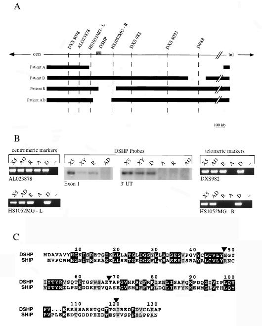
Figure 1.
Physical map of the XLP locus and identification of new genomic deletions disrupting DSHP. (A) Schematic representation of the XLP locus. The locus has been defined by a 3-Mb genomic deletion in patient A. Two new patients, R and AD, harbor deletions of 200 kb that span the DSHP gene, and are approximately 1 Mb centromeric to the previously reported 150 kb deletion in patient D. (B) Delineation of the borders of genomic deletions in patients R and AD, using PCR markers for centromeric and telomeric borders, and Southern blot analysis using probes from the 5′ and 3′ regions of DSHP. DSHP is entirely deleted in patient AD, whereas the centromeric breakpoint in patient R is between exons 1 and 2. (C) Comparison of the amino acid sequence of DSHP with the SH2 domain of SHIP. Identical amino acids are in black boxes, and conserved residues are in gray boxes. DSHP exon boundaries are indicated by arrowheads.

